
Intro
Discover the role of CT scans in heart health, including cardiac CT scans, coronary CT angiography, and heart scan diagnostics to detect cardiovascular diseases and conditions.
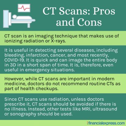
The importance of cardiovascular health cannot be overstated, as heart disease remains one of the leading causes of death worldwide. One of the most effective tools for diagnosing and monitoring heart conditions is the CT scan, specifically designed for cardiac imaging. This technology has revolutionized the field of cardiology, enabling doctors to detect potential issues before they become severe. With its high-resolution images and non-invasive procedure, CT scans for the heart have become an indispensable diagnostic tool. As we delve into the world of cardiac CT scans, it's essential to understand the benefits, working mechanisms, and steps involved in this life-saving procedure.
Cardiovascular diseases are often asymptomatic, making early detection crucial for effective treatment and prevention of complications. Traditional methods, such as electrocardiograms (ECGs) and stress tests, can provide valuable insights but may not offer the detailed visualization that a CT scan can. The CT scan's ability to capture high-resolution images of the heart and its surrounding blood vessels allows doctors to identify potential blockages, aneurysms, and other abnormalities that could lead to serious health issues. Furthermore, advancements in CT scan technology have significantly reduced radiation exposure, making it a safer option for patients.
The use of CT scans in cardiac imaging has opened up new avenues for the early detection and treatment of heart diseases. By providing clear and detailed images of the heart, doctors can now diagnose conditions that may have gone undetected with traditional methods. This early detection enables timely interventions, which can significantly improve patient outcomes. Moreover, the non-invasive nature of CT scans reduces the risk of complications associated with more invasive procedures, such as cardiac catheterization. As technology continues to evolve, the role of CT scans in cardiac care is expected to expand, offering new hope for patients and healthcare providers alike.
How CT Scans Work for Heart Imaging

CT scans use X-rays to create detailed cross-sectional images of the body, including the heart. In the context of cardiac imaging, CT scans are particularly useful for visualizing the coronary arteries, which supply blood to the heart muscle. The process involves the patient lying on a table that slides into a large, doughnut-shaped machine. The CT scanner rotates around the patient, taking X-ray images from different angles. These images are then reconstructed into detailed, three-dimensional pictures of the heart, allowing doctors to examine its structure and function from various perspectives.
The working mechanism of a CT scan for heart imaging involves several key steps:
- Preparation: The patient may be asked to change into a gown and remove any jewelry or metal objects that could interfere with the scan.
- Contrast Agent: A contrast agent, usually containing iodine, is injected into a vein to highlight the blood vessels and heart structures in the images.
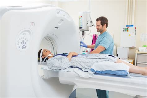
- Scan: The patient lies on the table, and the CT scanner rotates around them, capturing images from multiple angles.
- Image Reconstruction: The captured images are reconstructed into detailed, three-dimensional pictures of the heart.
- Analysis: A radiologist or cardiologist analyzes the images to identify any abnormalities or potential issues.
Benefits of CT Scans for Heart Imaging
The benefits of using CT scans for heart imaging are numerous and significant:
- High-Resolution Images: Provides detailed, three-dimensional pictures of the heart and its blood vessels.
- Non-Invasive: Reduces the risk of complications associated with more invasive diagnostic procedures.
- Early Detection: Enables the early detection of heart diseases, improving patient outcomes through timely interventions.
- Low Radiation Exposure: Modern CT scanners are designed to minimize radiation exposure, making them safer for patients.
- Wide Availability: CT scans are widely available in hospitals and diagnostic centers, making them accessible for a broad range of patients.
Steps Involved in a CT Scan for Heart Imaging
The process of undergoing a CT scan for heart imaging involves several steps:
- Scheduling and Preparation: The patient schedules the scan and may be advised on preparation, such as fasting or avoiding certain medications.
- Changing and Positioning: The patient changes into a gown and is positioned on the CT scanner table.
- Contrast Agent Administration: A contrast agent is injected to highlight the heart and blood vessels.
- Scanning: The CT scanner captures images of the heart from multiple angles.
- Image Analysis: A specialist analyzes the images for any signs of heart disease or abnormalities.
Practical Examples and Statistical Data
CT scans have been instrumental in diagnosing and managing heart diseases. For instance, a study found that the use of CT scans for coronary artery disease diagnosis resulted in a significant reduction in mortality rates compared to traditional diagnostic methods. Additionally, advancements in CT scan technology have improved image quality while reducing radiation exposure, making it a safer diagnostic tool for patients.
Some key statistical data include:
- Reduced Mortality Rates: Studies have shown that early detection and treatment facilitated by CT scans can reduce mortality rates from heart diseases.
- Increased Diagnostic Accuracy: CT scans offer high diagnostic accuracy for heart conditions, including coronary artery disease and cardiac arrhythmias.
- Wide Adoption: The use of CT scans for heart imaging has become widespread, reflecting their utility and effectiveness in clinical practice.
FAQs About CT Scans for Heart Imaging
What is a CT scan, and how does it work for heart imaging?
+A CT scan uses X-rays to create detailed images of the heart, allowing for the visualization of its structures and blood vessels. It's particularly useful for diagnosing coronary artery disease and other heart conditions.
Is a CT scan safe, and what are the potential risks?
+CT scans are generally safe, with modern machines designed to minimize radiation exposure. However, as with any medical procedure, there are potential risks, including allergic reactions to the contrast agent and radiation exposure.
How do I prepare for a CT scan for heart imaging?
+Preparation may include fasting, avoiding certain medications, and wearing comfortable clothing. Your healthcare provider will give you specific instructions based on your medical history and the type of scan you're undergoing.
As we conclude our exploration of CT scans for heart imaging, it's clear that this technology has revolutionized the field of cardiology. With its ability to provide detailed, high-resolution images of the heart and its blood vessels, CT scans have become an indispensable tool for diagnosing and managing heart diseases. Whether you're a healthcare professional or someone interested in learning more about cardiac health, understanding the role of CT scans can empower you to make informed decisions about your health or the health of your loved ones. We invite you to share your thoughts, ask questions, or explore further resources on this critical topic, as together, we can work towards a better understanding of heart health and the technologies that support it.