
Intro
Discover the importance of MRI of Lumbar Spine for diagnosing spinal issues, herniated discs, and nerve compression, using magnetic resonance imaging for accurate spinal cord and vertebrae assessment.
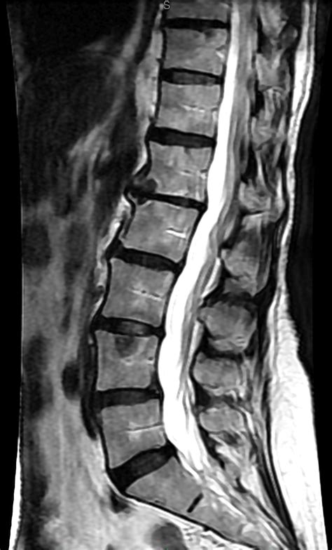
The lumbar spine, also known as the lower back, is a complex and vital part of the human body. It provides support, stability, and flexibility, allowing us to perform various daily activities. However, it is also prone to injuries, degenerative conditions, and other problems that can cause significant pain and discomfort. Magnetic Resonance Imaging (MRI) of the lumbar spine is a non-invasive and highly effective diagnostic tool that helps doctors to visualize the internal structures of the lower back, identify potential issues, and develop effective treatment plans.
The importance of MRI in diagnosing lumbar spine conditions cannot be overstated. It provides detailed images of the soft tissues, bones, and other structures, allowing doctors to diagnose a wide range of conditions, including herniated discs, spinal stenosis, spondylolisthesis, and spinal tumors. Moreover, MRI is a safe and painless procedure that does not involve the use of ionizing radiation, making it an attractive option for patients who require repeated imaging studies.
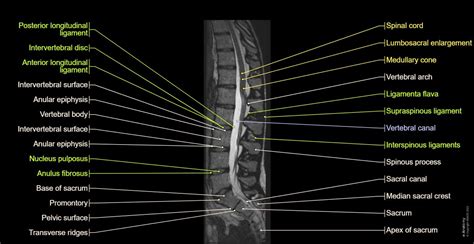
The lumbar spine is a complex and dynamic structure that consists of five vertebrae, intervertebral discs, facet joints, and various ligaments and muscles. Each of these components plays a crucial role in maintaining the integrity and function of the lower back. However, various factors, such as aging, trauma, and repetitive stress, can cause damage to these structures, leading to pain, stiffness, and limited mobility. MRI of the lumbar spine helps doctors to identify the underlying causes of these symptoms and develop targeted treatment plans that address the specific needs of each patient.
Introduction to MRI of Lumbar Spine
Benefits of MRI of Lumbar Spine
The benefits of MRI of the lumbar spine are numerous and well-documented. Some of the most significant advantages include: * High-resolution images of soft tissues, bones, and other structures * Non-invasive and painless procedure * No use of ionizing radiation * Ability to diagnose a wide range of conditions, including herniated discs, spinal stenosis, and spinal tumors * Helps doctors to develop targeted treatment plans that address the specific needs of each patientHow MRI of Lumbar Spine Works
Preparation for MRI of Lumbar Spine
To prepare for an MRI of the lumbar spine, patients should: * Avoid eating or drinking for at least 4-6 hours before the procedure * Remove any metal objects, such as jewelry, glasses, or clothing with metal fasteners * Inform their doctor about any medical conditions, such as pacemakers or implants * Wear comfortable clothing and avoid wearing anything with metalCommon Conditions Diagnosed with MRI of Lumbar Spine
Treatment Options for Lumbar Spine Conditions
Treatment options for lumbar spine conditions vary depending on the specific condition and severity of symptoms. Some common treatment options include: * Physical therapy: exercises and stretches to improve flexibility, strength, and range of motion * Medications: pain relievers, muscle relaxants, and anti-inflammatory medications to manage pain and inflammation * Surgery: surgical procedures to repair or replace damaged structures, such as herniated discs or spinal tumorsAdvantages and Disadvantages of MRI of Lumbar Spine
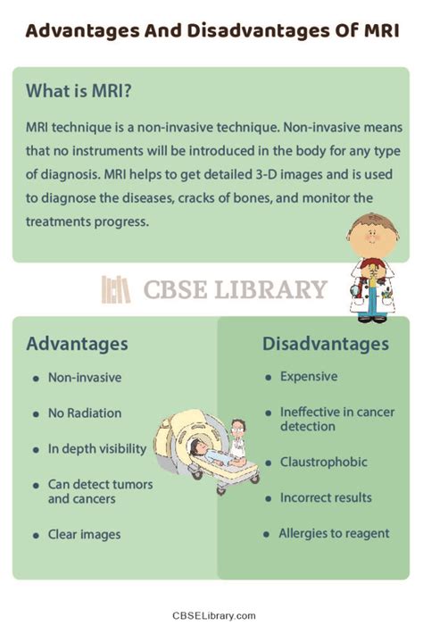
The disadvantages of MRI of the lumbar spine include:
- High cost
- Limited availability
- Claustrophobia or anxiety in some patients
- Requires specialized equipment and trained personnel
Future Developments in MRI of Lumbar Spine
Future developments in MRI of the lumbar spine include: * Improved image resolution and quality * Advanced imaging protocols and techniques * Increased availability and accessibility * Integration with other imaging modalities, such as CT and PET scansConclusion and Recommendations
Final Thoughts
MRI of the lumbar spine is a valuable diagnostic tool that can help patients to receive accurate diagnoses and effective treatment plans. We encourage patients to ask questions and seek a second opinion if necessary. By working together with their doctor, patients can take an active role in managing their lower back health and improving their overall quality of life.What is MRI of the lumbar spine?
+MRI of the lumbar spine is a non-invasive diagnostic tool that uses strong magnetic fields and radio waves to generate detailed images of the internal structures of the lower back.
What are the benefits of MRI of the lumbar spine?
+The benefits of MRI of the lumbar spine include high-resolution images of soft tissues, bones, and other structures, non-invasive and painless procedure, and no use of ionizing radiation.
What are the common conditions diagnosed with MRI of the lumbar spine?
+Common conditions diagnosed with MRI of the lumbar spine include herniated discs, spinal stenosis, spondylolisthesis, and spinal tumors.
We invite readers to share their thoughts and experiences with MRI of the lumbar spine in the comments section below. If you have any questions or concerns, please do not hesitate to ask. We also encourage readers to share this article with others who may benefit from this information. By working together, we can promote greater awareness and understanding of lower back health and the importance of accurate diagnosis and treatment.