
Intro
Discover the Tee medical procedure, a minimally invasive technique for treating varicose veins, spider veins, and vein disorders, using endovenous laser therapy and sclerotherapy for effective vein removal and relief.
The medical field is vast and intricate, with numerous procedures designed to diagnose, treat, and prevent a wide range of health conditions. Among these, the TEE medical procedure stands out for its critical role in assessing the heart's structure and function. Understanding what TEE is, its benefits, and how it works can provide valuable insights into cardiac health and the diagnostic tools available to healthcare professionals.
The importance of cardiac health cannot be overstated. The heart is a vital organ that pumps blood throughout the body, supplying tissues with oxygen and nutrients. Any condition that affects the heart's ability to perform this function can have significant implications for overall health. This is where diagnostic procedures like TEE come into play, offering a detailed look at the heart's condition and guiding treatment decisions.
For individuals facing heart-related issues, the prospect of undergoing a medical procedure can be daunting. Fear of the unknown often exacerbates anxiety, making it essential to educate patients about what to expect. The TEE procedure, while invasive, is generally safe and well-tolerated, providing crucial information that can lead to effective management and treatment of cardiac conditions. As technology advances and medical practices evolve, procedures like TEE continue to play a pivotal role in cardiovascular care.
Introduction to TEE Medical Procedure
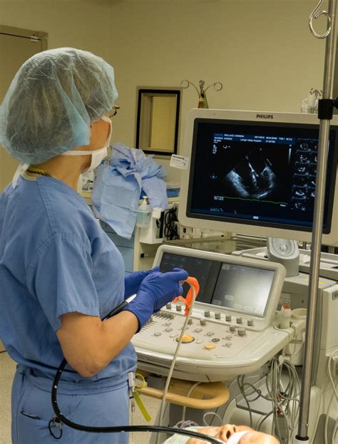
The TEE, or Transesophageal Echocardiogram, is a specialized ultrasound test used to produce detailed images of the heart and its blood vessels. Unlike a traditional echocardiogram, which is performed on the chest, TEE involves passing an ultrasound probe through the mouth and into the esophagus, which lies close to the heart. This proximity allows for higher quality images, especially of structures at the back of the heart, and can be particularly useful in certain situations where a standard echocardiogram may not provide sufficient detail.
Benefits of TEE
The benefits of TEE over other imaging techniques include its ability to provide clear, detailed pictures of the heart without the interference of the chest wall or lungs. This is especially beneficial for patients with certain conditions, such as obesity or chronic obstructive pulmonary disease (COPD), where traditional echocardiography might be less effective. Additionally, TEE can be used during surgical procedures to monitor the heart in real-time, allowing for immediate adjustments as needed.How TEE Works
The process of undergoing a TEE involves several steps. First, the patient is typically given sedation to help relax and reduce the gag reflex, making the procedure more comfortable. Next, a flexible tube (endoscope) with an ultrasound device on the end is gently guided through the mouth, down the throat, and into the esophagus. Once in place, the ultrasound device uses high-frequency sound waves to create detailed images of the heart, which are then displayed on a monitor for the healthcare team to analyze.
Preparation and Aftercare
Preparation for a TEE involves fasting for several hours before the procedure to ensure the stomach is empty, reducing the risk of complications. After the procedure, patients are monitored for a period, usually in a recovery area, until the sedation wears off. It's common for the throat to feel sore, and patients are advised not to eat or drink until their gag reflex returns to prevent choking. Instructions for post-procedure care, including diet and activity level, are provided by the healthcare team.Common Uses of TEE

TEE is utilized in various clinical scenarios, including the evaluation of heart valves, assessment of cardiac function in patients with heart failure, and examination for blood clots in the heart. It's also a valuable tool in the diagnosis of endocarditis, an infection of the heart valves, and in guiding certain interventional cardiac procedures. For patients undergoing heart surgery, TEE can provide critical real-time information, helping surgeons make informed decisions during the operation.
Risks and Complications
While generally safe, TEE is not without risks. Potential complications include bleeding, infection, and reactions to the sedation used during the procedure. There's also a small risk of damaging the esophagus or causing other internal injuries, although these are rare. Patients are closely monitored during and after the procedure to quickly identify and address any complications that may arise.Advancements in TEE Technology

Advancements in ultrasound technology continue to enhance the capabilities of TEE, allowing for even clearer images and more precise diagnoses. Three-dimensional (3D) echocardiography, for example, provides a more detailed view of the heart's anatomy than traditional 2D imaging, aiding in the assessment of complex heart defects and the planning of surgical interventions. Additionally, the development of smaller, more agile probes has made TEE more comfortable and accessible for patients.
Future Directions
The future of TEE holds promise for further innovation, with ongoing research focused on improving image quality, expanding the range of applications, and integrating TEE with other diagnostic and therapeutic modalities. As our understanding of cardiac disease and the tools available to diagnose and treat it evolve, procedures like TEE will continue to play a vital role in cardiovascular medicine, offering hope for better patient outcomes and improved quality of life.Conclusion and Next Steps

In conclusion, the TEE medical procedure represents a powerful diagnostic tool in the field of cardiology, offering detailed insights into the heart's structure and function. By understanding the benefits, process, and applications of TEE, patients and healthcare professionals alike can appreciate its value in managing cardiac health. As medical technology continues to advance, it's exciting to consider the potential future developments that will further enhance the capabilities of TEE and contribute to better patient care.
For those interested in learning more about TEE or who have questions about the procedure, we invite you to engage with our community. Share your experiences, ask questions, or simply stay updated on the latest advancements in cardiac care. Together, we can foster a deeper understanding of the heart and the many tools available to keep it healthy.
What is a TEE medical procedure?
+A TEE, or Transesophageal Echocardiogram, is a specialized ultrasound test that produces detailed images of the heart and its blood vessels by passing an ultrasound probe through the mouth and into the esophagus.
Why is TEE used over other imaging techniques?
+TEE provides clear, detailed pictures of the heart without interference from the chest wall or lungs, making it particularly useful for patients with certain conditions or during surgical procedures.
What are the risks and complications associated with TEE?
+Potential complications include bleeding, infection, reactions to sedation, and rare instances of esophageal damage. Patients are closely monitored to address any issues promptly.