
Intro
Discover how cystoscopy works with 5 key methods, utilizing endoscopy, urology, and medical imaging to diagnose urinary tract issues, bladder problems, and kidney stones, promoting urologic health and informed treatment decisions.
Cystoscopy is a medical procedure that allows doctors to visually examine the inside of the bladder and urethra. This procedure is crucial for diagnosing and treating various urinary tract problems, including bladder cancer, kidney stones, and urinary tract infections. The importance of cystoscopy lies in its ability to provide a clear and detailed view of the internal structures of the urinary system, enabling healthcare professionals to make accurate diagnoses and develop effective treatment plans. As a result, understanding how cystoscopy works is essential for patients who are about to undergo this procedure or for those who want to learn more about the diagnostic tools used in urology.
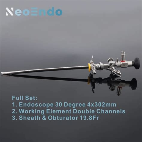
The process of cystoscopy involves the use of a cystoscope, which is a thin, flexible or rigid tube with a camera and light on the end. The cystoscope is inserted into the urethra and guided through to the bladder, allowing the doctor to see the internal structures on a monitor. This visual examination can reveal abnormalities such as tumors, stones, or inflammation, which may not be detectable through other diagnostic methods. The detailed visualization provided by cystoscopy is invaluable for both diagnostic purposes and for guiding therapeutic interventions, such as the removal of stones or the injection of medications directly into the bladder.
Cystoscopy is a relatively common procedure that is performed in both outpatient and inpatient settings. It is generally well-tolerated, although some patients may experience discomfort or mild pain during or after the procedure. The benefits of cystoscopy far outweigh the potential drawbacks, as it offers a direct and detailed view of the urinary tract, facilitating precise diagnoses and treatments. Moreover, advancements in technology have led to the development of more sophisticated cystoscopes, enhancing the clarity and resolution of the images obtained during the procedure. This technological progress has significantly improved the diagnostic and therapeutic capabilities of cystoscopy, making it an indispensable tool in the field of urology.
Introduction to Cystoscopy
Benefits of Cystoscopy

Working Mechanism of Cystoscopy
Steps Involved in Cystoscopy

Practical Examples and Statistical Data

Common Uses of Cystoscopy
Cystoscopy is commonly used for: - Diagnosing bladder cancer - Identifying urinary tract infections - Removing kidney stones - Injecting medications into the bladder - Performing biopsiesAdvancements in Cystoscopy Technology
Advancements in cystoscopy technology include the development of more flexible and thinner cystoscopes, high-definition cameras, and advanced imaging techniques such as narrow-band imaging (NBI) and photodynamic diagnosis (PDD). These advancements have improved the diagnostic accuracy of cystoscopy and expanded its therapeutic capabilities.FAQs About Cystoscopy
What is cystoscopy used for?
+Cystoscopy is used for the diagnosis and treatment of urinary tract problems, including bladder cancer, kidney stones, and urinary tract infections.
Is cystoscopy painful?
+Cystoscopy may cause some discomfort or mild pain, but anesthesia is used to minimize pain during the procedure.
How long does a cystoscopy procedure take?
+The length of the procedure can vary but typically takes between 15 to 30 minutes for a diagnostic cystoscopy.
In conclusion, cystoscopy is a vital diagnostic and therapeutic tool in the field of urology, offering a direct and detailed view of the urinary tract. Its benefits, working mechanisms, and steps involved make it an indispensable procedure for the management of various urinary tract disorders. As technology continues to advance, the capabilities and effectiveness of cystoscopy are expected to improve, further enhancing patient care and outcomes. We invite readers to share their experiences or ask questions about cystoscopy in the comments section below, and to consider sharing this informative article with others who may benefit from a deeper understanding of this important medical procedure.