
Intro
Discover the bone scan procedure, a diagnostic test using nuclear medicine to detect bone diseases, cancer, and injuries, utilizing radiotracers and imaging techniques for accurate diagnosis and treatment planning.
The bone scan procedure is a medical imaging technique used to diagnose and monitor various bone-related conditions, such as bone cancer, osteoporosis, and bone infections. This non-invasive test helps doctors to visualize the skeletal system and identify any abnormalities or areas of concern. In this article, we will delve into the details of the bone scan procedure, its benefits, and what patients can expect during the test.
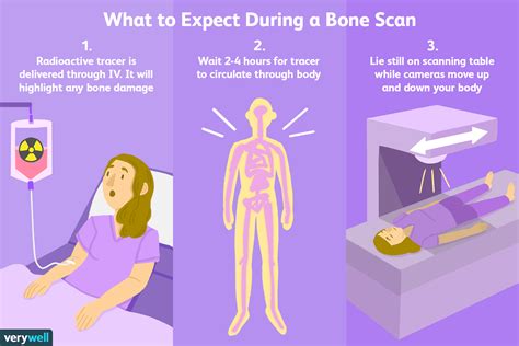
The bone scan procedure is typically used to diagnose conditions such as bone metastases, osteomyelitis, and Paget's disease. It can also be used to monitor the effectiveness of treatment for these conditions. The test is usually performed in a hospital or imaging center and takes about 30 minutes to an hour to complete. Patients are typically asked to arrive at the testing facility a few hours before the scheduled appointment time to allow for preparation and injection of a small amount of radioactive material.
The bone scan procedure involves the use of a small amount of radioactive material, usually technetium-99m, which is injected into the patient's bloodstream. This material accumulates in the bones and emits gamma rays, which are then detected by a special camera called a gamma camera. The camera takes pictures of the skeletal system, allowing doctors to visualize any areas of abnormal bone activity. The test is usually performed in two parts: the first part involves the injection of the radioactive material and a waiting period of about 2-3 hours, during which the material accumulates in the bones. The second part involves the actual scanning process, during which the patient lies on a table and the gamma camera takes pictures of the skeletal system.
Bone Scan Procedure Steps

Benefits of Bone Scan Procedure
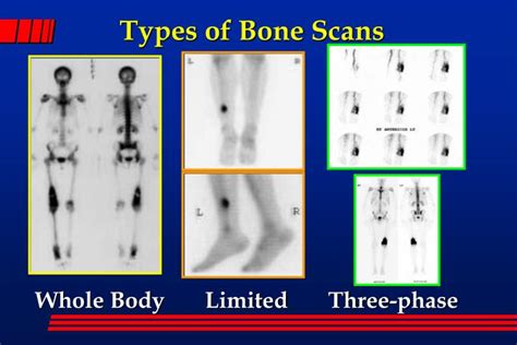
The bone scan procedure has several benefits, including its non-invasive nature, high sensitivity, and ability to detect abnormalities in the skeletal system. The test is also relatively painless and does not require any surgical incisions or insertion of instruments into the body. The bone scan procedure can also be used to monitor the effectiveness of treatment for various bone-related conditions, allowing doctors to adjust treatment plans as needed. Additionally, the test can help doctors to identify areas of abnormal bone activity, which can be useful in diagnosing conditions such as bone cancer or osteoporosis.Types of Bone Scans
Preparation for Bone Scan Procedure
Patients can prepare for the bone scan procedure by wearing comfortable clothing and removing any jewelry or clothing that may interfere with the test. They should also empty their bladder before the injection of the radioactive material. Patients may be asked to avoid eating or drinking for a few hours before the test, although this is not always necessary. It is also important for patients to inform their doctor about any medications they are taking, as some medications may interfere with the test. Women who are pregnant or breastfeeding should inform their doctor before undergoing the test, as the radioactive material may pass to the fetus or baby.Risks and Side Effects of Bone Scan Procedure

What to Expect After the Bone Scan Procedure
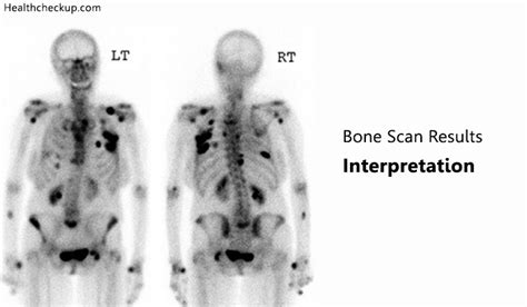
After the bone scan procedure, patients can usually return to their normal activities, although they may be asked to avoid close contact with pregnant women or young children for a few hours. The radioactive material used in the test is usually excreted in the urine and stool, and patients may be asked to flush the toilet twice after using it. Patients should also drink plenty of water to help flush out the radioactive material. The test results are usually available within a few hours, although they may take longer in some cases. Patients should discuss the test results with their doctor, who can explain the findings and recommend any further treatment or testing.Interpreting Bone Scan Results
Bone Scan Procedure Cost
The cost of the bone scan procedure can vary depending on the location, type of scan, and insurance coverage. On average, the cost of a whole-body bone scan can range from $1,000 to $3,000, although this can vary depending on the facility and location. Limited bone scans can cost less, ranging from $500 to $1,500. Patients should check with their insurance provider to see if the test is covered, as some insurance plans may not cover the full cost of the test.Bone Scan Procedure Alternatives
Conclusion and Next Steps
In conclusion, the bone scan procedure is a valuable diagnostic tool that can help doctors to diagnose and monitor various bone-related conditions. While the test is generally safe, there are some risks and side effects associated with the procedure. Patients should discuss the test results with their doctor, who can explain the findings and recommend any further treatment or testing. By understanding the bone scan procedure and its alternatives, patients can make informed decisions about their healthcare and take the next steps towards diagnosis and treatment.To learn more about the bone scan procedure and its applications, readers can explore the following resources:
- National Institutes of Health (NIH)
- American College of Radiology (ACR)
- Radiological Society of North America (RSNA)
We encourage readers to share their thoughts and experiences with the bone scan procedure in the comments section below. If you have any questions or concerns about the test, please do not hesitate to ask.
What is a bone scan procedure?
+A bone scan procedure is a medical imaging test that uses a small amount of radioactive material to visualize the skeletal system and diagnose various bone-related conditions.
What are the benefits of a bone scan procedure?
+The benefits of a bone scan procedure include its non-invasive nature, high sensitivity, and ability to detect abnormalities in the skeletal system. The test can also be used to monitor the effectiveness of treatment for various bone-related conditions.
What are the risks and side effects of a bone scan procedure?
+The risks and side effects of a bone scan procedure include allergic reactions, fatigue, nausea, and dizziness. In rare cases, the test can cause more serious side effects, such as anaphylaxis or radiation exposure.