
Intro
Understand normal chest X-ray results, including lung fields, cardiac silhouette, and mediastinum. Learn to interpret radiographic findings, diagnose conditions, and identify abnormalities with related terms like pulmonary health, cardiorespiratory systems, and thoracic imaging.
A chest X-ray is a crucial diagnostic tool used by medical professionals to evaluate the lungs, heart, and chest cavity. It is a non-invasive and painless procedure that helps doctors diagnose and monitor various medical conditions, such as pneumonia, lung cancer, and heart disease. Understanding normal chest X-ray results is essential for both medical professionals and patients to ensure accurate diagnosis and treatment. In this article, we will delve into the world of chest X-rays, exploring what a normal result entails, how the procedure works, and what to expect during the diagnostic process.
The importance of chest X-rays cannot be overstated, as they provide valuable insights into the inner workings of the chest cavity. By examining the lungs, heart, and surrounding tissues, doctors can identify potential issues before they become severe. A normal chest X-ray result is a significant indicator of overall health, and it can help alleviate concerns and worries for patients. However, it is crucial to understand that a normal result does not necessarily mean that a person is completely healthy, as some conditions may not be visible on an X-ray.
Chest X-rays have been a cornerstone of medical diagnosis for decades, and their significance extends beyond the medical community. Patients who undergo chest X-rays often have questions and concerns about the procedure, the results, and what they mean. By explaining normal chest X-ray results in detail, we aim to empower patients with knowledge, reducing anxiety and uncertainty. Whether you are a medical professional or a patient, understanding the intricacies of chest X-rays is essential for making informed decisions about your health.
Introduction to Chest X-Rays

A chest X-ray is a type of radiographic examination that uses low-level radiation to produce images of the chest cavity. The procedure involves exposing the chest to a small amount of X-ray radiation, which passes through the body and onto a digital detector or film. The resulting image provides a detailed view of the lungs, heart, and surrounding tissues, allowing doctors to diagnose and monitor various medical conditions.
How Chest X-Rays Work
The process of taking a chest X-ray is relatively straightforward. The patient stands or sits in front of the X-ray machine, and the technician positions the machine to capture the desired view. The most common views are the posterior-anterior (PA) view, which shows the chest from the back to the front, and the lateral view, which shows the chest from the side. The technician then exposes the chest to a small amount of X-ray radiation, which passes through the body and onto the digital detector or film.Understanding Normal Chest X-Ray Results
A normal chest X-ray result indicates that the lungs, heart, and surrounding tissues appear healthy and free from any significant abnormalities. The radiologist or doctor examines the X-ray image for signs of disease, injury, or other conditions that may affect the chest cavity. A normal result typically means that the:
- Lungs are clear and free from any signs of disease or injury
- Heart is normal in size and shape
- Chest cavity is free from any signs of fluid or air accumulation
- Bones and soft tissues appear healthy and intact
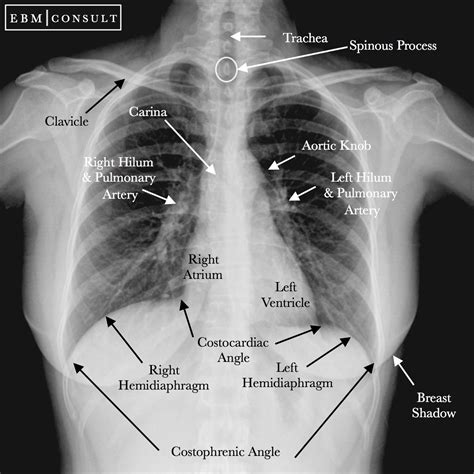
Components of a Normal Chest X-Ray Result
A normal chest X-ray result includes several key components, which are essential for accurate diagnosis and treatment. These components include:- Lung fields: The lung fields should be clear and free from any signs of disease or injury, such as nodules, masses, or consolidations.
- Cardiac silhouette: The cardiac silhouette should be normal in size and shape, indicating a healthy heart.
- Mediastinum: The mediastinum, which is the area between the lungs, should be free from any signs of widening or masses.
- Pleura: The pleura, which is the membrane surrounding the lungs, should be intact and free from any signs of fluid or air accumulation.
- Bones and soft tissues: The bones and soft tissues, including the ribs, vertebrae, and muscles, should appear healthy and intact.
Abnormal Chest X-Ray Results
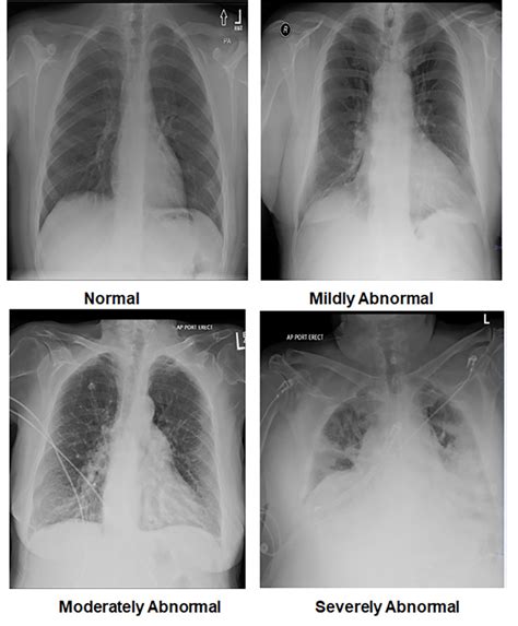
An abnormal chest X-ray result indicates that there is an issue with the lungs, heart, or surrounding tissues. Abnormal results can be caused by a variety of conditions, including pneumonia, lung cancer, heart disease, and injuries. Some common signs of abnormality on a chest X-ray include:
- Nodules or masses in the lung fields
- Consolidations or infiltrates in the lung fields
- Cardiac enlargement or abnormal shape
- Widening of the mediastinum
- Pleural effusion or pneumothorax
Causes of Abnormal Chest X-Ray Results
Abnormal chest X-ray results can be caused by a variety of conditions, including:- Infections, such as pneumonia or tuberculosis
- Cancer, such as lung cancer or lymphoma
- Heart disease, such as cardiomegaly or coronary artery disease
- Injuries, such as rib fractures or pneumothorax
- Congenital conditions, such as cystic fibrosis or congenital heart disease
Limitations of Chest X-Rays
While chest X-rays are a valuable diagnostic tool, they have several limitations. These limitations include:
- Limited sensitivity: Chest X-rays may not detect all types of lung disease or injury, particularly in the early stages.
- Limited specificity: Chest X-rays may not provide a definitive diagnosis, and further testing may be necessary to confirm a diagnosis.
- Radiation exposure: Chest X-rays involve exposure to low-level radiation, which can be a concern for patients who require frequent X-rays.
Alternatives to Chest X-Rays
In some cases, alternatives to chest X-rays may be necessary or preferred. These alternatives include:- Computed tomography (CT) scans: CT scans provide more detailed images of the chest cavity and can detect smaller lesions or abnormalities.
- Magnetic resonance imaging (MRI): MRI provides detailed images of the chest cavity and can detect abnormalities in the soft tissues.
- Ultrasound: Ultrasound can be used to evaluate the lungs and surrounding tissues, particularly in cases where X-rays are not feasible.
Conclusion and Next Steps

In conclusion, understanding normal chest X-ray results is essential for both medical professionals and patients. By recognizing the components of a normal result and the limitations of chest X-rays, patients can better understand their diagnosis and treatment options. If you have undergone a chest X-ray and have questions or concerns about your results, it is essential to discuss them with your doctor or radiologist. They can provide you with more information and guidance on the next steps in your care.
We invite you to share your thoughts and experiences with chest X-rays in the comments below. Have you undergone a chest X-ray and received a normal or abnormal result? What were your concerns, and how did you navigate the diagnostic process? Your stories and insights can help others who are going through similar experiences.
What is a normal chest X-ray result?
+A normal chest X-ray result indicates that the lungs, heart, and surrounding tissues appear healthy and free from any significant abnormalities.
What are the components of a normal chest X-ray result?
+The components of a normal chest X-ray result include clear lung fields, a normal cardiac silhouette, a normal mediastinum, intact pleura, and healthy bones and soft tissues.
What are the limitations of chest X-rays?
+The limitations of chest X-rays include limited sensitivity, limited specificity, and radiation exposure.