
Intro
The abdominal region of the human body is a complex and vital area, housing essential organs such as the stomach, liver, kidneys, and intestines. When it comes to diagnosing and treating conditions affecting these organs, medical professionals often rely on imaging tests like computed tomography (CT) scans. A CT scan of the abdomen is a non-invasive and highly effective diagnostic tool that provides detailed cross-sectional images of the abdominal organs and tissues. In this article, we will delve into the world of CT scans, exploring their benefits, working mechanisms, and the steps involved in undergoing an abdominal CT scan.
CT scans have revolutionized the field of medical imaging, offering a rapid and accurate means of diagnosing a wide range of abdominal conditions, from appendicitis and kidney stones to liver disease and cancer. The high-resolution images produced by CT scans enable doctors to pinpoint the exact location and extent of abnormalities, allowing for timely and targeted treatment. Moreover, CT scans are often used to monitor the progression of diseases and the effectiveness of treatments, making them an indispensable tool in modern medicine.
The importance of CT scans cannot be overstated, particularly in emergency situations where every minute counts. For instance, a CT scan can quickly diagnose internal injuries or bleeding in trauma patients, guiding surgeons and emergency responders in their decision-making. Similarly, in cases of suspected appendicitis or intestinal obstruction, a CT scan can confirm the diagnosis and help determine the best course of treatment. As we explore the world of abdominal CT scans, it becomes clear that this technology has transformed the way medical professionals approach diagnosis and treatment, saving countless lives and improving patient outcomes.
How CT Scans Work
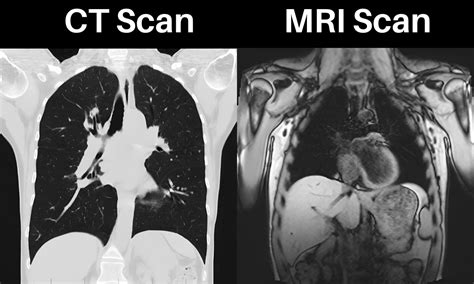
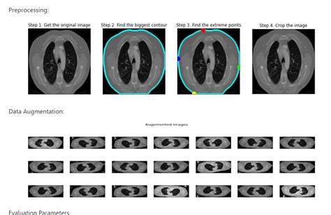
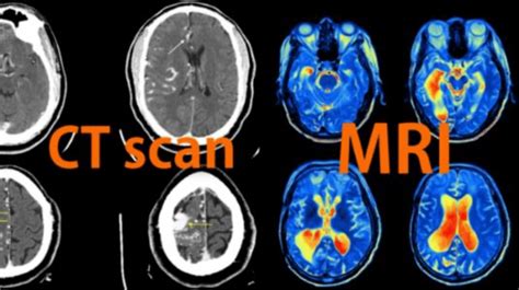
CT scans utilize a combination of X-rays and computer technology to produce detailed images of the body's internal structures. During a CT scan, the patient lies on a table that slides into a large, doughnut-shaped machine. The machine is equipped with X-ray detectors and emitters, which rotate around the patient, capturing multiple images from different angles. These images are then reconstructed by a computer, creating a three-dimensional picture of the abdominal organs and tissues. The resulting images can be viewed on a monitor, allowing doctors to zoom in, rotate, and manipulate the images to gain a better understanding of the patient's condition.
The working mechanism of CT scans is based on the principle of differential absorption of X-rays by various tissues. Different tissues, such as bone, muscle, and fat, absorb X-rays to varying degrees, allowing the CT scanner to distinguish between them. By detecting the amount of X-ray absorption, the scanner can create detailed images of the body's internal structures, including organs, blood vessels, and tissues. This technology has been refined over the years, with modern CT scanners capable of producing high-resolution images with unprecedented speed and accuracy.
Preparation and Procedure

Before undergoing a CT scan, patients may be required to prepare in various ways, depending on the specific examination and their individual needs. This may include fasting for a certain period, avoiding certain medications, or wearing comfortable clothing. In some cases, patients may be asked to drink a contrast agent, such as barium or iodine, to help highlight specific areas of the body. The contrast agent can be administered orally or intravenously, depending on the type of examination and the patient's medical history.
The CT scan procedure itself is relatively quick and painless, typically taking between 10 to 30 minutes to complete. The patient lies on the table, which slides into the CT scanner, and remains still during the examination. The scanner's X-ray detectors and emitters rotate around the patient, capturing images from multiple angles. The patient may be asked to hold their breath or remain still for short periods to ensure clear images. In some cases, the CT scanner may be equipped with advanced features, such as cardiac gating or respiratory tracking, to improve image quality and reduce motion artifacts.
Benefits and Risks
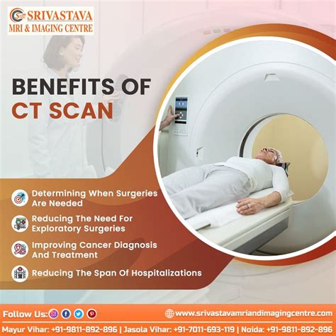
CT scans offer numerous benefits, including rapid diagnosis, high-resolution images, and minimal invasiveness. The detailed images produced by CT scans enable doctors to diagnose a wide range of conditions, from acute appendicitis to chronic liver disease. Additionally, CT scans can help guide biopsies, tumor treatments, and other minimally invasive procedures, reducing the need for surgical intervention.
However, like any medical imaging test, CT scans also carry certain risks and limitations. The primary concern is radiation exposure, as CT scans use X-rays to produce images. While the radiation doses used in CT scans are generally considered safe, repeated exposure can increase the risk of cancer and other health problems. Moreover, CT scans may not be suitable for pregnant women or individuals with certain medical conditions, such as kidney disease or allergies to contrast agents.
Common Applications
CT scans have a wide range of applications in medical imaging, including:
- Diagnosing abdominal conditions, such as appendicitis, diverticulitis, and intestinal obstruction
- Monitoring cancer treatment and detecting tumor recurrence
- Guiding biopsies and minimally invasive procedures
- Evaluating liver and kidney function
- Detecting kidney stones and gallstones
- Assessing abdominal trauma and internal injuries
CT scans can also be used to monitor the progression of diseases, such as liver cirrhosis or chronic kidney disease, and to evaluate the effectiveness of treatments. Additionally, CT scans can help identify potential health risks, such as atherosclerosis or abdominal aortic aneurysms, allowing for early intervention and prevention.
CT Scan Types
There are several types of CT scans, each with its own unique characteristics and applications. These include:
- Conventional CT scans: The most common type of CT scan, which uses a single X-ray source and detector
- Spiral CT scans: A type of CT scan that uses a spiral or helical motion to capture images, allowing for faster scanning and higher resolution
- High-speed CT scans: A type of CT scan that uses advanced technology to capture images at high speeds, reducing scan times and improving image quality
- Low-dose CT scans: A type of CT scan that uses lower radiation doses, reducing the risk of radiation exposure and making it suitable for patients who require repeated scans
Each type of CT scan has its own advantages and disadvantages, and the choice of scan depends on the specific medical condition, patient needs, and scanner availability.
Contrast Agents
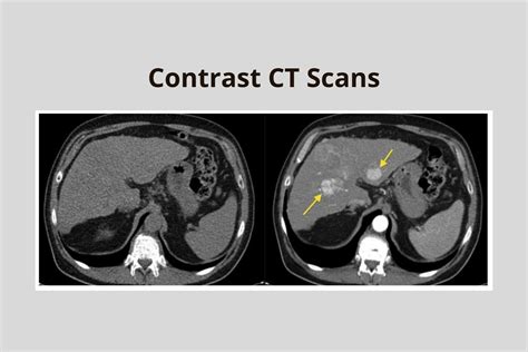
Contrast agents are substances used to enhance the visibility of certain areas or structures during a CT scan. These agents can be administered orally, intravenously, or rectally, depending on the type of examination and patient needs. Common contrast agents used in CT scans include:
- Barium sulfate: A contrast agent used to visualize the gastrointestinal tract
- Iodine-based agents: Contrast agents used to visualize blood vessels, kidneys, and other organs
- Gadolinium-based agents: Contrast agents used to visualize the liver, pancreas, and other organs
Contrast agents can help highlight specific areas of the body, making it easier to diagnose conditions and monitor treatment effectiveness. However, contrast agents can also carry risks, such as allergic reactions or kidney damage, and should be used judiciously and under medical supervision.
CT Scan Results
CT scan results are typically interpreted by a radiologist, who analyzes the images to diagnose conditions, detect abnormalities, and monitor treatment effectiveness. The results may be communicated to the patient's doctor, who will then discuss the findings and recommend further treatment or testing.
CT scan results can be categorized into several types, including:
- Normal: The CT scan shows no abnormalities or signs of disease
- Abnormal: The CT scan shows signs of disease or abnormalities, such as tumors, inflammation, or damage
- Inconclusive: The CT scan results are unclear or require further testing to confirm a diagnosis
In some cases, CT scan results may require additional testing or procedures to confirm a diagnosis or monitor treatment effectiveness. Patients should discuss their results with their doctor and ask questions to understand their condition and treatment options.
Future Developments
The field of CT scanning is constantly evolving, with advances in technology and techniques improving image quality, reducing radiation exposure, and expanding applications. Some future developments in CT scanning include:
- Artificial intelligence: The use of AI algorithms to improve image analysis, detect abnormalities, and predict patient outcomes
- Machine learning: The use of machine learning techniques to develop personalized treatment plans and predict treatment responses
- Low-dose CT scans: The development of low-dose CT scans that reduce radiation exposure while maintaining image quality
These advancements are expected to further improve the diagnostic accuracy and therapeutic effectiveness of CT scans, making them an even more valuable tool in modern medicine.
What is a CT scan, and how does it work?
+A CT scan is a medical imaging test that uses X-rays and computer technology to produce detailed images of the body's internal structures. It works by detecting the amount of X-ray absorption by different tissues, allowing the scanner to create detailed images of organs, blood vessels, and tissues.
What are the benefits and risks of CT scans?
+CT scans offer numerous benefits, including rapid diagnosis, high-resolution images, and minimal invasiveness. However, they also carry risks, such as radiation exposure, allergic reactions to contrast agents, and kidney damage.
How do I prepare for a CT scan, and what can I expect during the procedure?
+To prepare for a CT scan, you may need to fast, avoid certain medications, or wear comfortable clothing. During the procedure, you will lie on a table that slides into the CT scanner, and remain still while the scanner captures images. You may be asked to hold your breath or remain still for short periods to ensure clear images.
What are the common applications of CT scans, and how are they used in medical imaging?
+CT scans have a wide range of applications, including diagnosing abdominal conditions, monitoring cancer treatment, guiding biopsies, and evaluating liver and kidney function. They are used to produce detailed images of internal structures, allowing doctors to diagnose conditions, detect abnormalities, and monitor treatment effectiveness.
What are the different types of CT scans, and how do they differ from each other?
+There are several types of CT scans, including conventional, spiral, high-speed, and low-dose CT scans. Each type has its own unique characteristics and applications, and the choice of scan depends on the specific medical condition, patient needs, and scanner availability.
As we conclude our exploration of CT scans, it is clear that this technology has revolutionized the field of medical imaging, offering a rapid, accurate, and non-invasive means of diagnosing and treating a wide range of conditions. Whether you are a medical professional, a patient, or simply interested in learning more about CT scans, we hope this article has provided you with a comprehensive understanding of this valuable tool. We invite you to share your thoughts, ask questions, or seek further information on this topic, and we look forward to continuing the conversation.