
Intro
Discover the Cxr medical abbreviation meaning, a crucial radiology term. Learn about chest X-ray diagnostics, medical imaging, and radiographic procedures.
The medical field is filled with abbreviations and acronyms that can be confusing for those not familiar with them. One such abbreviation is CXR, which is commonly used in medical settings. Understanding the meaning of CXR is essential for both medical professionals and patients to ensure effective communication and proper care. In this article, we will delve into the world of medical abbreviations, focusing on CXR, its meaning, and its significance in the medical field.
CXR is an abbreviation that stands for Chest X-Ray, a diagnostic imaging test used to examine the lungs, heart, and other structures within the chest cavity. Chest X-Rays are a crucial tool for diagnosing and monitoring various medical conditions, including respiratory diseases, heart conditions, and lung injuries. The test involves exposing the chest to a small amount of ionizing radiation, which produces images of the internal structures. These images can help healthcare providers identify abnormalities, such as tumors, cysts, or fractures, and develop appropriate treatment plans.
The importance of understanding medical abbreviations like CXR cannot be overstated. In medical settings, clear and precise communication is critical for providing quality care. Misinterpretation of abbreviations can lead to misunderstandings, misdiagnoses, or even adverse events. Therefore, it is essential for healthcare professionals, patients, and families to be familiar with common medical abbreviations, including CXR.
Introduction to Chest X-Rays
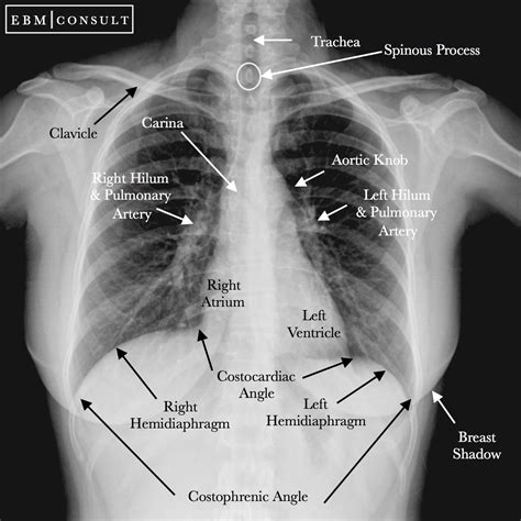
Chest X-Rays are a non-invasive and relatively quick procedure, typically taking only a few minutes to complete. The test is usually performed in a radiology department or a dedicated X-Ray suite. During the procedure, the patient stands or sits in front of an X-Ray machine, and the technician positions the machine to capture the desired images. The X-Ray machine emits a low-level radiation beam, which passes through the chest and onto a digital detector or film. The resulting images are then reviewed by a radiologist or other healthcare provider to identify any abnormalities or conditions.
Benefits of Chest X-Rays
Chest X-Rays offer several benefits, including:
- Non-invasive and relatively painless procedure
- Quick and easy to perform
- Low radiation exposure
- Can help diagnose a wide range of medical conditions
- Inexpensive compared to other imaging tests
The benefits of Chest X-Rays make them a valuable diagnostic tool in various medical settings, from emergency departments to outpatient clinics.
Common Uses of Chest X-Rays
Chest X-Rays are commonly used to: * Diagnose respiratory diseases, such as pneumonia or bronchitis * Evaluate the size and shape of the heart * Detect lung injuries or conditions, such as pulmonary embolism or pneumothorax * Monitor the progression of medical conditions, such as cystic fibrosis or lung cancer * Guide medical procedures, such as biopsies or drainage of fluid collectionsHow Chest X-Rays Work

Chest X-Rays work by using ionizing radiation to produce images of the internal structures within the chest cavity. The X-Ray machine emits a beam of radiation, which passes through the chest and onto a digital detector or film. The resulting images are then reviewed by a radiologist or other healthcare provider to identify any abnormalities or conditions.
The process of producing a Chest X-Ray image involves several steps:
- The patient is positioned in front of the X-Ray machine.
- The X-Ray machine is adjusted to the correct settings.
- The X-Ray beam is emitted, passing through the chest.
- The resulting image is captured on a digital detector or film.
- The image is reviewed by a radiologist or other healthcare provider.
Risks and Limitations of Chest X-Rays
While Chest X-Rays are generally safe, there are some risks and limitations to consider: * Radiation exposure: Chest X-Rays involve exposure to ionizing radiation, which can increase the risk of cancer or other health problems. * Limited diagnostic capability: Chest X-Rays may not detect all medical conditions, particularly those that do not involve the lungs or heart. * Inconclusive results: In some cases, Chest X-Ray results may be inconclusive, requiring additional testing or evaluation.Preparing for a Chest X-Ray

Preparing for a Chest X-Ray is relatively straightforward. Patients are typically asked to:
- Remove any jewelry or clothing that may interfere with the X-Ray beam
- Wear a hospital gown or other comfortable clothing
- Stand or sit in front of the X-Ray machine
- Remain still during the procedure to ensure clear images
In some cases, patients may be asked to hold their breath or perform specific breathing exercises to help capture the desired images.
Interpreting Chest X-Ray Results
Interpreting Chest X-Ray results requires specialized training and expertise. Radiologists and other healthcare providers review the images to identify any abnormalities or conditions. The results may be reported as: * Normal: The Chest X-Ray appears normal, with no signs of disease or injury. * Abnormal: The Chest X-Ray shows signs of disease or injury, such as tumors, cysts, or fractures. * Inconclusive: The Chest X-Ray results are unclear or require additional testing or evaluation.Conclusion and Next Steps

In conclusion, understanding the meaning and significance of CXR is essential for effective communication and proper care in medical settings. Chest X-Rays are a valuable diagnostic tool, offering a non-invasive and relatively quick procedure for examining the lungs, heart, and other structures within the chest cavity. By familiarizing themselves with CXR and other medical abbreviations, healthcare professionals, patients, and families can work together to provide quality care and improve health outcomes.
We invite you to share your thoughts and experiences with Chest X-Rays in the comments below. Have you had a Chest X-Ray recently? What was your experience like? Do you have any questions or concerns about the procedure? We encourage you to ask questions, share your story, or provide feedback on this article.
What is a Chest X-Ray used for?
+A Chest X-Ray is used to diagnose and monitor various medical conditions, including respiratory diseases, heart conditions, and lung injuries.
How long does a Chest X-Ray take?
+A Chest X-Ray typically takes only a few minutes to complete.
Are Chest X-Rays safe?
+Chest X-Rays involve exposure to ionizing radiation, which can increase the risk of cancer or other health problems. However, the benefits of the procedure often outweigh the risks.
How do I prepare for a Chest X-Ray?
+Patients are typically asked to remove any jewelry or clothing that may interfere with the X-Ray beam, wear a hospital gown or other comfortable clothing, and stand or sit in front of the X-Ray machine.
What are the risks and limitations of Chest X-Rays?
+Chest X-Rays involve radiation exposure, may not detect all medical conditions, and can produce inconclusive results. However, the benefits of the procedure often outweigh the risks, and additional testing or evaluation can be performed if necessary.