
Intro
Improve X-ray interpretation with 5 hand X-ray tips, including positioning, alignment, and diagnostic techniques for accurate bone and joint analysis, fracture detection, and osteoporosis assessment.
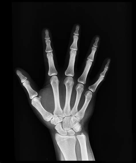
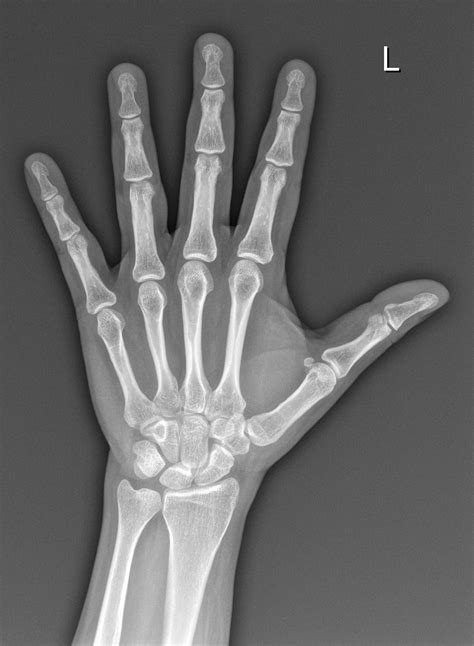
The importance of hand X-rays cannot be overstated, particularly in the fields of medicine and health diagnostics. Hand X-rays are a crucial diagnostic tool used to evaluate the bones and joints in the hands, helping doctors to diagnose and treat a wide range of conditions, from fractures and dislocations to arthritis and bone tumors. With the advancement of technology, hand X-rays have become more sophisticated, providing clearer images and more accurate diagnoses. In this article, we will delve into the world of hand X-rays, exploring their benefits, working mechanisms, and providing valuable tips for patients undergoing this procedure.
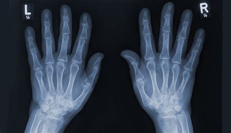
Hand X-rays are a non-invasive and relatively quick procedure, typically taking only a few minutes to complete. During the procedure, the patient's hand is placed on a flat surface, and an X-ray machine is positioned above it. The machine emits low-level radiation, which passes through the hand, capturing images of the bones and joints. These images are then displayed on a screen, allowing doctors to examine the hand in detail and diagnose any potential issues. The use of hand X-rays has revolutionized the field of orthopedics, enabling doctors to provide more accurate diagnoses and effective treatments.
The benefits of hand X-rays are numerous, ranging from diagnosing fractures and dislocations to monitoring the progression of arthritis and other conditions. Hand X-rays can also help doctors to identify any abnormalities in the bones or joints, such as bone spurs or cysts, which can cause pain and discomfort. Furthermore, hand X-rays can be used to monitor the effectiveness of treatments, such as physical therapy or medication, and to track the progression of conditions over time. With the increasing demand for hand X-rays, it is essential to understand the procedure, its benefits, and how to prepare for it.
Understanding Hand X-Ray Procedures
Preparation and Safety Precautions
To ensure a safe and successful hand X-ray procedure, patients must follow certain preparation and safety precautions. These include removing any jewelry or clothing that may interfere with the X-ray machine, avoiding movement during the procedure, and informing the doctor of any medical conditions or implants. Additionally, patients who are pregnant or breastfeeding should inform their doctor before undergoing a hand X-ray, as the radiation may pose a risk to the fetus or baby.Benefits and Risks of Hand X-Rays

Common Uses of Hand X-Rays
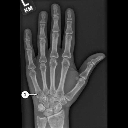
Hand X-rays are commonly used to diagnose and treat a wide range of conditions, including fractures, dislocations, arthritis, and bone tumors. They can also be used to monitor the progression of conditions over time and to track the effectiveness of treatments. Some of the most common uses of hand X-rays include: * Diagnosing fractures and dislocations * Monitoring the progression of arthritis and other conditions * Identifying abnormalities in the bones or joints * Tracking the effectiveness of treatments * Monitoring the progression of conditions over timeHand X-Ray Tips for Patients
What to Expect During a Hand X-Ray
During a hand X-ray, patients can expect to undergo a series of steps to ensure accurate and clear images. These include: * Removing any jewelry or clothing that may interfere with the X-ray machine * Placing the hand on a flat surface * Positioning the X-ray machine above the hand * Emitting low-level radiation to capture images of the bones and joints * Displaying the images on a screen for the doctor to examineInterpreting Hand X-Ray Results
Common Hand X-Ray Findings
Some common hand X-ray findings include: * Fractures or dislocations * Arthritis or other joint conditions * Bone spurs or cysts * Tumors or other abnormalities * Degenerative changes or wear and tearAdvances in Hand X-Ray Technology
Future Developments in Hand X-Ray Technology
Future developments in hand X-ray technology are expected to further improve the accuracy and effectiveness of the procedure. These may include: * Improved digital X-ray machines * Advanced computer-aided detection software * Increased use of artificial intelligence and machine learning * Development of new X-ray technologies, such as 3D imagingWhat is a hand X-ray, and how does it work?
+A hand X-ray is a medical imaging procedure that uses low-level radiation to capture images of the bones and joints in the hand. It works by emitting X-rays through the hand, which are then displayed on a screen for the doctor to examine.
What are the benefits of hand X-rays?
+The benefits of hand X-rays include diagnosing fractures and dislocations, monitoring the progression of arthritis and other conditions, identifying abnormalities in the bones or joints, and tracking the effectiveness of treatments.
What are the risks associated with hand X-rays?
+The risks associated with hand X-rays include exposure to radiation and potential allergic reactions to the X-ray dye. Patients should discuss these risks with their doctor before undergoing a hand X-ray.
In summary, hand X-rays are a valuable diagnostic tool used to evaluate the bones and joints in the hands. By understanding the benefits, working mechanisms, and risks associated with hand X-rays, patients can make informed decisions about their care. We invite you to share your thoughts and experiences with hand X-rays in the comments below. If you found this article helpful, please share it with others who may benefit from this information. Additionally, we encourage you to take action and consult with your doctor if you have any concerns about your hand health. By working together, we can promote better health and well-being for everyone.