
Intro
Discover the ins and outs of MRI of the head, including brain scans, neuroimaging, and diagnostic procedures, to understand how magnetic resonance imaging helps diagnose head injuries, brain disorders, and neurological conditions.
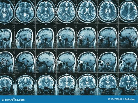
Magnetic Resonance Imaging (MRI) of the head is a diagnostic imaging technique that provides detailed images of the brain and its surrounding structures. This non-invasive procedure is crucial in diagnosing and monitoring various neurological conditions, such as tumors, strokes, and multiple sclerosis. The importance of MRI of the head cannot be overstated, as it enables healthcare professionals to visualize the brain's anatomy and detect potential abnormalities with unparalleled precision.
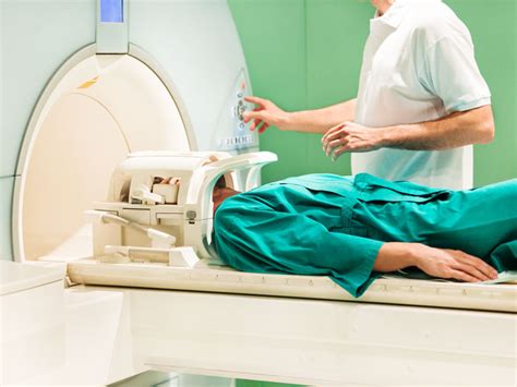
The process of undergoing an MRI of the head is relatively straightforward. Patients are typically asked to lie on a table that slides into a large, cylindrical machine. The machine uses a strong magnetic field and radio waves to generate images of the brain, which are then reconstructed by a computer. The entire procedure usually takes between 15 to 90 minutes, depending on the specific requirements of the scan. During this time, patients may be required to hold still and follow breathing instructions to ensure that the images are clear and accurate.
The significance of MRI of the head extends beyond its diagnostic capabilities. It also plays a vital role in treatment planning and monitoring. For instance, MRI can help surgeons pinpoint the location of tumors, allowing for more precise and effective surgical interventions. Additionally, MRI can be used to monitor the progression of neurological conditions, enabling healthcare professionals to adjust treatment plans accordingly. As medical technology continues to evolve, the applications of MRI of the head are likely to expand, further solidifying its position as a cornerstone of neurological diagnosis and treatment.
How MRI Of The Head Works

Key Components Of MRI Of The Head
The key components of MRI of the head include the magnetic field, radio waves, and computer systems. The magnetic field is responsible for aligning the hydrogen atoms in the body, while the radio waves disturb these atoms, causing them to emit signals. The computer systems reconstruct these signals into detailed images of the brain, which are then interpreted by healthcare professionals.Benefits Of MRI Of The Head

Common Applications Of MRI Of The Head
The common applications of MRI of the head include: * Diagnosing neurological conditions: MRI of the head is used to diagnose various neurological conditions, such as tumors, strokes, and multiple sclerosis. * Monitoring treatment: MRI of the head is used to monitor the effectiveness of treatment plans and adjust them accordingly. * Surgical planning: MRI of the head is used to plan surgical interventions, allowing surgeons to pinpoint the location of tumors and other abnormalities.Preparation For MRI Of The Head
What To Expect During The Scan
During the scan, patients can expect to: * Lie on a table: Patients will be asked to lie on a table that slides into the MRI machine. * Hold still: Patients will be required to hold still and follow breathing instructions to ensure that the images are clear and accurate. * Hear loud noises: The MRI machine can be loud, and patients may hear knocking or banging noises during the scan.Risks And Side Effects Of MRI Of The Head
Contrast Agents Used In MRI Of The Head
Contrast agents are substances used to enhance the visibility of certain structures or abnormalities in the brain. The most common contrast agent used in MRI of the head is gadolinium, which is generally safe and well-tolerated. However, some patients may be allergic to gadolinium, and alternative contrast agents may be used.Interpreting MRI Of The Head Results
Common Abnormalities Detected By MRI Of The Head
Some common abnormalities detected by MRI of the head include: * Tumors: MRI of the head can detect tumors, including brain cancer, meningiomas, and acoustic neuromas. * Strokes: MRI of the head can detect strokes, including ischemic and hemorrhagic strokes. * Multiple sclerosis: MRI of the head can detect multiple sclerosis, a chronic condition that affects the central nervous system.Advances In MRI Of The Head Technology
Future Directions For MRI Of The Head
The future of MRI of the head is exciting and promising. Some potential developments include: * Improved resolution: Advances in MRI technology may lead to higher-resolution images, enabling healthcare professionals to detect smaller abnormalities. * Increased sensitivity: Improved sensitivity may enable healthcare professionals to detect abnormalities earlier, leading to better treatment outcomes. * Personalized medicine: MRI of the head may be used to develop personalized treatment plans, tailored to individual patients' needs and conditions.What is MRI of the head used for?
+MRI of the head is used to diagnose and monitor various neurological conditions, including tumors, strokes, and multiple sclerosis.
Is MRI of the head safe?
+MRI of the head is generally a safe procedure, but there are some potential risks and side effects to be aware of, including claustrophobia, allergic reactions, and metal object interference.
How long does an MRI of the head take?
+The length of an MRI of the head can vary, but it typically takes between 15 to 90 minutes, depending on the specific requirements of the scan.
What are the benefits of MRI of the head?
+The benefits of MRI of the head include high-resolution images, non-invasive procedure, no radiation, and diagnostic accuracy.
Can I have an MRI of the head if I have a pacemaker?
+It's generally not recommended to have an MRI of the head if you have a pacemaker, as the strong magnetic field can interfere with the device. However, some MRI machines are designed to be compatible with certain pacemakers, so it's best to consult with your healthcare provider.
In conclusion, MRI of the head is a powerful diagnostic tool that has revolutionized the field of neurology. Its ability to provide high-resolution images of the brain and its surrounding structures has enabled healthcare professionals to diagnose and monitor various neurological conditions with unprecedented accuracy. As technology continues to evolve, the applications of MRI of the head are likely to expand, further solidifying its position as a cornerstone of neurological diagnosis and treatment. If you have any questions or concerns about MRI of the head, we encourage you to consult with your healthcare provider or leave a comment below. Share this article with others who may benefit from this information, and stay tuned for future updates on the latest advances in MRI technology.