Intro
Discover the Tee Echo Procedure Explained, a diagnostic echo test using transesophageal echocardiography (TEE) for cardiac imaging, providing detailed heart views and assessing cardiovascular health through ultrasound technology.
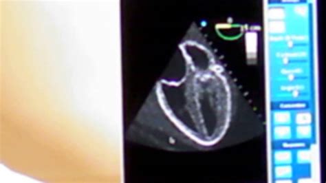
The Tee Echo procedure, also known as Transesophageal Echocardiography, is a medical imaging test used to produce high-quality images of the heart and its blood vessels. This procedure is essential for diagnosing and monitoring various heart conditions, including heart valve problems, coronary artery disease, and cardiac tumors. The importance of the Tee Echo procedure lies in its ability to provide detailed images of the heart's structure and function, allowing healthcare professionals to make accurate diagnoses and develop effective treatment plans.
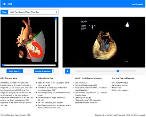
The Tee Echo procedure is a valuable diagnostic tool that has revolutionized the field of cardiology. By using high-frequency sound waves, this procedure can produce detailed images of the heart's anatomy, including the heart chambers, valves, and blood vessels. This information is crucial for identifying potential problems, such as blood clots, heart valve defects, and cardiac tumors. Moreover, the Tee Echo procedure can help healthcare professionals monitor the effectiveness of treatments and make adjustments as needed.
The significance of the Tee Echo procedure extends beyond its diagnostic capabilities. This procedure is also used to guide minimally invasive surgical procedures, such as cardiac catheterization and percutaneous valve repair. By providing real-time images of the heart, the Tee Echo procedure enables healthcare professionals to navigate the heart's anatomy with precision, reducing the risk of complications and improving patient outcomes. As a result, the Tee Echo procedure has become an essential tool in the management of heart disease, and its applications continue to expand as technology advances.
What is the Tee Echo Procedure?

Preparation for the Tee Echo Procedure
To prepare for the Tee Echo procedure, patients are typically asked to fast for several hours before the test. This is to reduce the risk of complications, such as aspiration, and to ensure that the stomach is empty. Patients may also be asked to stop taking certain medications, such as blood thinners, before the procedure. Additionally, patients should inform their healthcare provider about any allergies or sensitivities they may have, as well as any medical conditions that may affect the procedure.Benefits of the Tee Echo Procedure

Risks and Complications of the Tee Echo Procedure
While the Tee Echo procedure is generally safe, there are some risks and complications associated with it. These include: * Bleeding or perforation of the esophagus * Infection * Allergic reactions to the sedation or other medications * Cardiac arrhythmias or other heart problems * Respiratory problems, such as aspiration or pneumoniaApplications of the Tee Echo Procedure

Limitations of the Tee Echo Procedure
While the Tee Echo procedure is a valuable diagnostic tool, it does have some limitations. These include: * Limited view of the heart's anterior structures, such as the right ventricle * Difficulty visualizing the heart's posterior structures, such as the left atrium * Limited ability to evaluate the heart's coronary arteries * May not be suitable for patients with certain medical conditions, such as esophageal strictures or bleeding disordersFuture Directions of the Tee Echo Procedure

Conclusion and Recommendations
In conclusion, the Tee Echo procedure is a valuable diagnostic tool that has revolutionized the field of cardiology. Its high-quality images, minimally invasive nature, and real-time capabilities make it an essential tool for diagnosing and monitoring heart disease. While it does have some limitations, the Tee Echo procedure is continually evolving, with advances in technology and technique expanding its applications. As such, it is recommended that healthcare professionals consider the Tee Echo procedure as a first-line diagnostic tool for patients with suspected heart disease.What is the Tee Echo procedure used for?
+The Tee Echo procedure is used to produce high-quality images of the heart and its blood vessels, allowing healthcare professionals to diagnose and monitor various heart conditions.
Is the Tee Echo procedure painful?
+The Tee Echo procedure is typically performed under sedation, which minimizes discomfort. However, some patients may experience mild discomfort or throat irritation after the procedure.
What are the risks and complications of the Tee Echo procedure?
+The Tee Echo procedure is generally safe, but there are some risks and complications associated with it, including bleeding or perforation of the esophagus, infection, and cardiac arrhythmias.
We hope this article has provided you with a comprehensive understanding of the Tee Echo procedure and its applications. If you have any further questions or would like to share your experiences with the Tee Echo procedure, please do not hesitate to comment below. Additionally, if you found this article informative, please share it with others who may benefit from this information.
