
Intro
Discover 5 expert tips for a successful Transesophageal Echocardiogram, including preparation, procedure, and interpretation, to ensure accurate heart function diagnosis and effective cardiac care management.
The transesophageal echocardiogram, or TEE, is a specialized medical imaging test used to produce high-quality images of the heart and its blood vessels. This diagnostic tool is essential for assessing various heart conditions, including valve problems, blood clots, and tumors. As a non-invasive procedure, TEE has become a crucial component of cardiovascular care, providing valuable insights that guide treatment decisions. With its ability to deliver detailed, real-time images, TEE has revolutionized the field of cardiology, enabling healthcare professionals to diagnose and manage heart diseases more effectively.
The importance of TEE lies in its capacity to offer a more comprehensive view of the heart's structure and function compared to traditional echocardiograms. By inserting an ultrasound probe into the esophagus, which is located close to the heart, TEE can capture high-resolution images that are not possible with other imaging modalities. This proximity to the heart allows for clearer visualization of cardiac structures, making it an invaluable tool for diagnosing complex heart conditions. Moreover, TEE is a relatively safe procedure with minimal risks, making it an attractive option for patients who require detailed cardiac assessments.
As medical technology continues to advance, the role of TEE in cardiovascular care is likely to expand. With ongoing research and development, new applications for TEE are emerging, further solidifying its position as a critical diagnostic tool. Whether used for diagnostic purposes, guiding surgical interventions, or monitoring treatment outcomes, TEE has become an indispensable asset in the management of heart diseases. Its impact on patient care is significant, enabling healthcare providers to make informed decisions that improve outcomes and enhance the quality of life for individuals with cardiac conditions.
Understanding the Transesophageal Echocardiogram Procedure
Benefits of the Transesophageal Echocardiogram
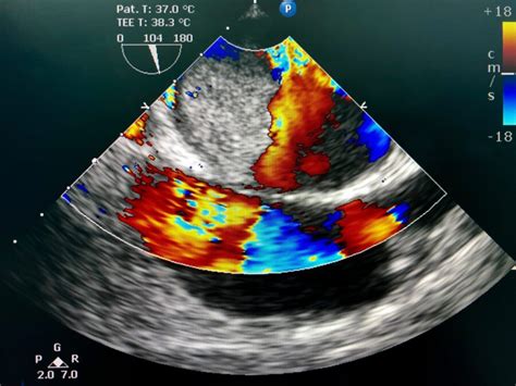
The benefits of TEE are numerous, including its ability to provide high-quality images of the heart's structures, such as the valves, chambers, and blood vessels. This detailed visualization enables healthcare providers to diagnose a wide range of heart conditions, from mitral valve prolapse to atrial septal defects. Additionally, TEE can be used to guide certain cardiac procedures, such as catheter-based interventions, and to monitor the effectiveness of treatments over time. Its non-invasive nature also makes it an attractive option for patients who may not be candidates for more invasive diagnostic tests.Preparing for a Transesophageal Echocardiogram

What to Expect During the Procedure
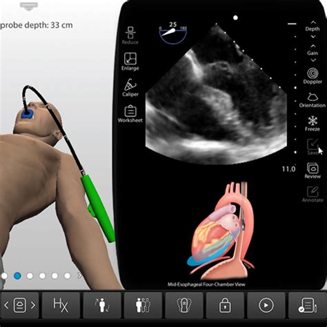
During the TEE procedure, the patient will be asked to lie on their side on an examination table. The healthcare provider will then insert the ultrasound probe into the mouth and guide it down the esophagus. This may cause some discomfort, but the local anesthetic and sedative should help minimize any pain or anxiety. Once the probe is in position, the healthcare provider will begin the imaging process, capturing detailed pictures of the heart and its structures. The procedure typically takes about 30 minutes to an hour to complete, although this may vary depending on the individual case.Risks and Complications of Transesophageal Echocardiogram
Interpreting Transesophageal Echocardiogram Results
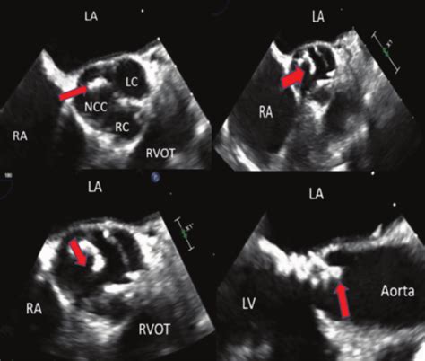
Interpreting TEE results requires specialized training and expertise. The images captured during the procedure are carefully reviewed by a cardiologist or other qualified healthcare professional to identify any abnormalities or signs of heart disease. The results may indicate the presence of conditions such as valve disease, cardiac tumors, or blood clots, among others. Based on the findings, the healthcare provider will develop a treatment plan tailored to the individual's needs, which may involve medication, surgery, or other interventions. In some cases, additional testing may be necessary to confirm the diagnosis or monitor the effectiveness of treatment.Advances in Transesophageal Echocardiogram Technology

Future Directions for Transesophageal Echocardiogram
As research and development continue to push the boundaries of TEE technology, we can expect to see new and innovative applications emerge. One area of focus is the development of smaller, more flexible probes that can be used in a wider range of patients, including children and individuals with certain anatomical limitations. Another area of exploration is the use of TEE in guiding minimally invasive surgical procedures, such as transcatheter valve replacements. With its ability to provide real-time imaging, TEE has the potential to play a critical role in these procedures, enhancing their safety and effectiveness.Transesophageal Echocardiogram in Clinical Practice

Best Practices for Transesophageal Echocardiogram
To ensure the safe and effective use of TEE, healthcare providers should follow established best practices. This includes proper patient preparation, careful probe insertion and manipulation, and meticulous image interpretation. Additionally, healthcare providers should be aware of potential complications and take steps to minimize risks. Regular training and education are also essential to stay up-to-date with the latest advancements in TEE technology and techniques. By adhering to these best practices, healthcare providers can optimize the benefits of TEE while minimizing its risks.Conclusion and Future Perspectives
Final Thoughts on Transesophageal Echocardiogram
As we look to the future, it's essential to recognize the significance of TEE in shaping the landscape of cardiovascular care. By leveraging its capabilities, healthcare providers can make more informed decisions, develop more effective treatment plans, and improve patient outcomes. As research and development continue to push the boundaries of TEE technology, we can expect to see new and exciting advancements emerge. Whether you're a healthcare provider, a patient, or simply interested in learning more about this innovative technology, it's clear that the transesophageal echocardiogram will remain a vital tool in the diagnosis and treatment of heart diseases for years to come.We invite you to share your thoughts, experiences, or questions about the transesophageal echocardiogram in the comments below. Your engagement is valuable to us, and we look forward to hearing your perspectives on this critical diagnostic tool. Additionally, if you found this article informative, please consider sharing it with others who may benefit from learning more about TEE.
What is a transesophageal echocardiogram?
+A transesophageal echocardiogram, or TEE, is a medical imaging test used to produce high-quality images of the heart and its blood vessels.
How is a transesophageal echocardiogram performed?
+The TEE procedure involves inserting an ultrasound probe into the esophagus, which is guided down to the desired position behind the heart to capture detailed images.
What are the benefits of a transesophageal echocardiogram?
+The benefits of TEE include its ability to provide high-quality images of the heart, guide certain cardiac procedures, and monitor the effectiveness of treatments over time.
Are there any risks associated with a transesophageal echocardiogram?
+While TEE is generally considered safe, potential risks include bleeding or perforation of the esophagus, throat discomfort, and allergic reactions to the sedative used during the procedure.
How long does a transesophageal echocardiogram procedure take?
+The TEE procedure typically takes about 30 minutes to an hour to complete, although this may vary depending on the individual case and the specific requirements of the test.