Intro
Discover what a bone scan is, a medical imaging test using nuclear medicine to diagnose bone diseases, injuries, and cancer, utilizing LSI keywords like osteoporosis, bone cancer, and nuclear scans.
Bone scans are a vital diagnostic tool used in the medical field to detect and monitor various bone-related conditions. The importance of bone scans lies in their ability to provide detailed information about the skeletal system, helping doctors diagnose and treat conditions such as bone cancer, osteoporosis, and bone infections. As we delve into the world of bone scans, it's essential to understand the significance of this diagnostic technique and how it can impact patient care.
The human skeletal system is a complex and dynamic structure, comprising 206 bones that provide support, protection, and mobility. However, bones can be affected by various diseases and conditions, making diagnosis and treatment challenging. Bone scans have revolutionized the field of orthopedics and oncology, enabling doctors to visualize the skeletal system in greater detail. By using bone scans, medical professionals can identify abnormalities, track disease progression, and monitor treatment efficacy.
The use of bone scans has become increasingly prevalent in recent years, driven by advances in medical technology and the growing need for accurate diagnoses. As the population ages, the incidence of bone-related conditions is expected to rise, making bone scans an essential tool in the diagnostic arsenal. Moreover, bone scans can help reduce the risk of misdiagnosis, ensuring that patients receive appropriate treatment and care. With the help of bone scans, doctors can develop personalized treatment plans, improving patient outcomes and quality of life.
What is a Bone Scan?

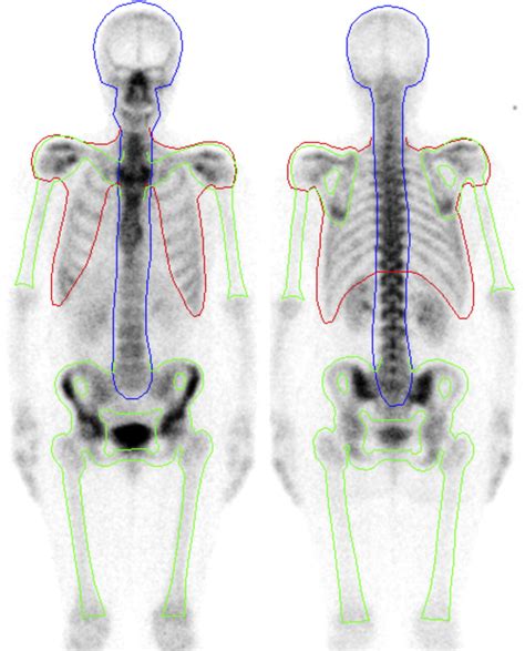
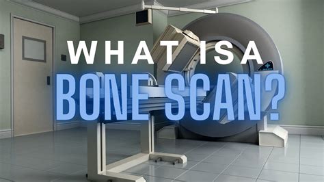
A bone scan, also known as a bone scintigraphy, is a diagnostic imaging test that uses small amounts of radioactive material to visualize the skeletal system. The test involves injecting a radioactive tracer into the bloodstream, which accumulates in the bones, allowing doctors to visualize the skeletal system using specialized cameras. The resulting images provide valuable information about bone metabolism, helping doctors diagnose and monitor various bone-related conditions.
How Does a Bone Scan Work?
The bone scan process typically involves the following steps: * The patient is injected with a small amount of radioactive tracer, usually technetium-99m-methyl diphosphonate (Tc-99m MDP). * The tracer accumulates in the bones, binding to hydroxyapatite, a mineral found in bone tissue. * The patient is then asked to wait for a period, usually 2-3 hours, to allow the tracer to accumulate in the bones. * The patient is then scanned using a gamma camera, which detects the radiation emitted by the tracer. * The resulting images are reconstructed to create a detailed picture of the skeletal system.Types of Bone Scans

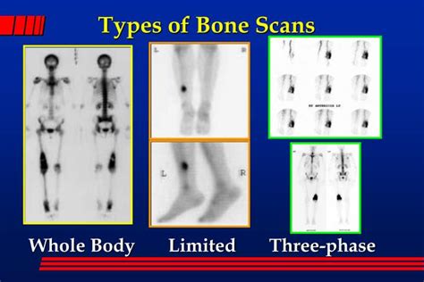
There are several types of bone scans, each with its own specific indications and applications. Some of the most common types of bone scans include:
- Planar bone scan: This is the most common type of bone scan, which provides a two-dimensional image of the skeletal system.
- Single-photon emission computed tomography (SPECT) bone scan: This type of scan provides a three-dimensional image of the skeletal system, offering greater detail and accuracy.
- Positron emission tomography (PET) bone scan: This type of scan uses a different type of radioactive tracer and provides a more detailed image of the skeletal system.
- Whole-body bone scan: This type of scan involves scanning the entire body, providing a comprehensive view of the skeletal system.
Indications for Bone Scans
Bone scans are used to diagnose and monitor a wide range of bone-related conditions, including: * **Bone cancer**: Bone scans can help diagnose and stage bone cancer, as well as monitor treatment efficacy. * **Osteoporosis**: Bone scans can help diagnose and monitor osteoporosis, a condition characterized by weakened bones. * **Bone infections**: Bone scans can help diagnose and monitor bone infections, such as osteomyelitis. * **Bone fractures**: Bone scans can help diagnose and monitor bone fractures, particularly in cases where conventional imaging modalities are inconclusive.Benefits of Bone Scans

Bone scans offer several benefits, including:
- High sensitivity: Bone scans are highly sensitive, allowing doctors to detect abnormalities in the skeletal system.
- Non-invasive: Bone scans are non-invasive, making them a safe and comfortable diagnostic procedure.
- Low radiation exposure: Bone scans involve low radiation exposure, making them a safe diagnostic procedure.
- Comprehensive view: Bone scans provide a comprehensive view of the skeletal system, allowing doctors to diagnose and monitor conditions that affect multiple bones.
Risks and Side Effects
While bone scans are generally safe, there are some risks and side effects to consider: * **Radiation exposure**: Bone scans involve radiation exposure, which can increase the risk of cancer and genetic damage. * **Allergic reactions**: Some patients may experience allergic reactions to the radioactive tracer. * **Pain and discomfort**: Some patients may experience pain and discomfort during the injection procedure.Preparation and Aftercare

To prepare for a bone scan, patients should:
- Avoid caffeine and nicotine: Patients should avoid caffeine and nicotine for at least 24 hours before the test.
- Wear comfortable clothing: Patients should wear comfortable clothing and avoid wearing metal jewelry or accessories.
- Remove metal objects: Patients should remove any metal objects, such as glasses or dentures, before the test. After the test, patients should:
- Drink plenty of water: Patients should drink plenty of water to help flush out the radioactive tracer.
- Avoid close contact: Patients should avoid close contact with others, particularly pregnant women and young children, for at least 24 hours after the test.
Interpretation of Results
The results of a bone scan are typically interpreted by a radiologist or a nuclear medicine specialist. The results may show: * **Normal bone activity**: Normal bone activity is indicated by a uniform distribution of the radioactive tracer. * **Abnormal bone activity**: Abnormal bone activity is indicated by an increased or decreased uptake of the radioactive tracer. * **Bone lesions**: Bone lesions, such as tumors or fractures, may appear as areas of increased or decreased uptake.Future Directions

The future of bone scans looks promising, with advances in technology and techniques expected to improve diagnostic accuracy and patient outcomes. Some of the future directions include:
- Hybrid imaging: Hybrid imaging techniques, such as SPECT/CT or PET/CT, are expected to provide more detailed and accurate images of the skeletal system.
- New tracers: New tracers, such as those targeting specific bone receptors, are being developed to improve diagnostic accuracy and specificity.
- Artificial intelligence: Artificial intelligence and machine learning algorithms are being developed to improve image analysis and interpretation.
Conclusion and Recommendations
In conclusion, bone scans are a vital diagnostic tool used in the medical field to detect and monitor various bone-related conditions. The benefits of bone scans, including high sensitivity, non-invasiveness, and low radiation exposure, make them a safe and effective diagnostic procedure. However, patients should be aware of the risks and side effects, and follow preparation and aftercare instructions carefully. As the field of bone scans continues to evolve, we can expect to see improvements in diagnostic accuracy and patient outcomes.What is a bone scan used for?
+A bone scan is used to diagnose and monitor various bone-related conditions, such as bone cancer, osteoporosis, and bone infections.
How long does a bone scan take?
+A bone scan typically takes 2-3 hours, including preparation and scanning time.
Are bone scans safe?
+Bone scans are generally safe, but they do involve radiation exposure, which can increase the risk of cancer and genetic damage.
Can I eat and drink before a bone scan?
+Yes, you can eat and drink before a bone scan, but you should avoid caffeine and nicotine for at least 24 hours before the test.
How often can I have a bone scan?
+The frequency of bone scans depends on the specific condition being monitored and the doctor's recommendations.
We hope this article has provided you with a comprehensive understanding of bone scans and their role in diagnosing and monitoring bone-related conditions. If you have any further questions or concerns, please don't hesitate to comment below or share this article with others.
