
Intro
Discover the ultimate Computed Tomography Brain Scan Guide, covering CT scan procedures, brain imaging techniques, and neurological diagnostic methods, to understand brain health and detect conditions like stroke and tumors.
The human brain is a complex and fascinating organ, and understanding its functions and structures is crucial for diagnosing and treating various neurological conditions. One of the most effective diagnostic tools for visualizing the brain is the Computed Tomography (CT) brain scan. This non-invasive medical imaging technique uses X-rays and computer technology to produce detailed cross-sectional images of the brain, allowing healthcare professionals to identify abnormalities, injuries, and diseases. In this article, we will delve into the world of CT brain scans, exploring their benefits, working mechanisms, and applications in neurology.
The importance of CT brain scans cannot be overstated, as they have revolutionized the field of neurology and saved countless lives. By providing high-resolution images of the brain, CT scans enable doctors to diagnose conditions such as strokes, tumors, and traumatic brain injuries with greater accuracy and speed. Furthermore, CT scans are widely available, relatively quick, and generally less expensive than other imaging modalities like Magnetic Resonance Imaging (MRI). As a result, CT brain scans have become an essential tool in emergency medicine, neurosurgery, and neurology.
The process of undergoing a CT brain scan is relatively straightforward and painless. Patients are typically asked to lie on a table that slides into a large, doughnut-shaped machine, which houses the X-ray tube and detectors. The X-ray tube rotates around the patient's head, emitting narrow beams of X-rays that pass through the brain and are detected by the sensors. The resulting data is then reconstructed into detailed images of the brain using sophisticated computer algorithms. The entire procedure usually takes between 10-30 minutes, depending on the type of scan and the patient's condition.
How CT Brain Scans Work
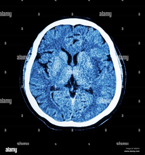
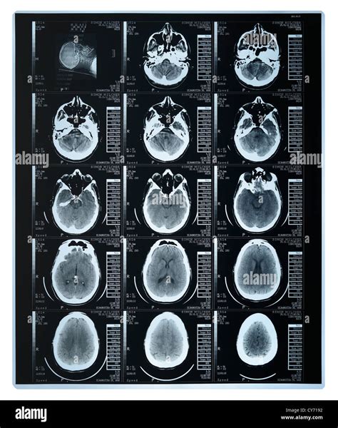
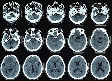
CT brain scans work by using X-rays to produce detailed images of the brain's internal structures. The X-ray tube emits a narrow beam of X-rays that passes through the patient's head, and the detectors measure the amount of X-rays that are absorbed or scattered by the brain tissue. The resulting data is then reconstructed into images using computer algorithms, which take into account the density of the brain tissue and the X-ray absorption coefficients. The images are typically displayed in a series of cross-sectional slices, allowing doctors to visualize the brain's anatomy and identify any abnormalities.
Types of CT Brain Scans
There are several types of CT brain scans, each with its own specific applications and advantages. Some of the most common types of CT brain scans include: * Non-contrast CT scans: These scans use X-rays alone to produce images of the brain and are often used to diagnose conditions such as strokes, traumatic brain injuries, and cerebral hemorrhages. * Contrast-enhanced CT scans: These scans use a contrast agent, such as iodine or barium, to highlight specific areas of the brain and are often used to diagnose conditions such as tumors, infections, and vascular malformations. * CT angiography: This type of scan uses a contrast agent to visualize the blood vessels in the brain and is often used to diagnose conditions such as aneurysms, arteriovenous malformations, and vascular stenosis. * CT perfusion: This type of scan uses a contrast agent to measure blood flow and perfusion in the brain and is often used to diagnose conditions such as strokes and cerebral vasospasm.Benefits of CT Brain Scans
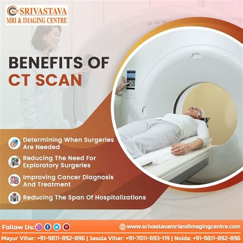
CT brain scans offer several benefits, including:
- High-resolution images: CT scans produce detailed images of the brain, allowing doctors to visualize small structures and abnormalities.
- Fast and convenient: CT scans are relatively quick, taking between 10-30 minutes to complete, and are widely available in hospitals and imaging centers.
- Non-invasive: CT scans are non-invasive, meaning that they do not require surgery or the insertion of instruments into the brain.
- Cost-effective: CT scans are generally less expensive than other imaging modalities, such as MRI or PET scans.
- Wide range of applications: CT scans can be used to diagnose a wide range of conditions, including strokes, tumors, traumatic brain injuries, and vascular malformations.
Applications of CT Brain Scans
CT brain scans have a wide range of applications in neurology and neurosurgery, including: * Diagnosing strokes and cerebral vasospasm * Detecting tumors, such as glioblastomas and meningiomas * Visualizing traumatic brain injuries, such as concussions and subdural hematomas * Identifying vascular malformations, such as aneurysms and arteriovenous malformations * Guiding neurosurgical procedures, such as tumor resections and aneurysm clippingPreparing for a CT Brain Scan
Preparing for a CT brain scan is relatively straightforward, but there are several steps that patients can take to ensure that the procedure is safe and effective. Some of the key steps include:
- Removing jewelry and clothing that contains metal
- Avoiding food and drink for several hours before the scan
- Informing the doctor about any medical conditions, such as diabetes or kidney disease
- Avoiding contrast agents if there is a history of allergy or sensitivity
- Following the doctor's instructions for any specific preparations, such as drinking a contrast agent or undergoing a bowel prep
Risks and Side Effects
While CT brain scans are generally safe, there are several risks and side effects that patients should be aware of. Some of the key risks and side effects include: * Radiation exposure: CT scans use X-rays, which can expose patients to ionizing radiation. * Contrast agent reactions: Some patients may be allergic or sensitive to the contrast agents used in CT scans. * Kidney damage: The contrast agents used in CT scans can cause kidney damage in patients with pre-existing kidney disease. * Claustrophobia: Some patients may experience claustrophobia or anxiety during the CT scan.Interpreting CT Brain Scan Results
Interpreting CT brain scan results requires a high degree of expertise and training, as the images can be complex and nuanced. Some of the key factors that doctors consider when interpreting CT brain scan results include:
- The presence of abnormalities, such as tumors or vascular malformations
- The size and location of any abnormalities
- The density of the brain tissue and the X-ray absorption coefficients
- The presence of any secondary signs, such as edema or mass effect
Common Abnormalities
Some of the most common abnormalities that can be detected on CT brain scans include: * Strokes and cerebral vasospasm * Tumors, such as glioblastomas and meningiomas * Traumatic brain injuries, such as concussions and subdural hematomas * Vascular malformations, such as aneurysms and arteriovenous malformations * Infections, such as meningitis and encephalitisAdvances in CT Brain Scan Technology
CT brain scan technology is constantly evolving, with advances in detector design, X-ray tube technology, and computer algorithms. Some of the key advances in CT brain scan technology include:
- High-resolution detectors: New detector designs have improved the resolution of CT scans, allowing for more detailed images of the brain.
- Dual-energy CT: This technology uses two different X-ray energies to produce images of the brain, allowing for better differentiation between different types of tissue.
- Spectral CT: This technology uses multiple X-ray energies to produce images of the brain, allowing for better characterization of tissue composition and function.
Future Directions
The future of CT brain scan technology is exciting and promising, with several new developments on the horizon. Some of the key future directions include: * Artificial intelligence: AI algorithms are being developed to help interpret CT brain scan results, reducing the time and expertise required for diagnosis. * Machine learning: Machine learning algorithms are being developed to help improve the accuracy and speed of CT brain scan interpretations. * Personalized medicine: CT brain scans are being used to develop personalized treatment plans, tailored to the individual patient's needs and condition.What is a CT brain scan?
+A CT brain scan is a medical imaging test that uses X-rays and computer technology to produce detailed cross-sectional images of the brain.
What are the benefits of CT brain scans?
+CT brain scans offer several benefits, including high-resolution images, fast and convenient scanning, non-invasive procedures, and cost-effectiveness.
What are the risks and side effects of CT brain scans?
+The risks and side effects of CT brain scans include radiation exposure, contrast agent reactions, kidney damage, and claustrophobia.
How do I prepare for a CT brain scan?
+To prepare for a CT brain scan, patients should remove jewelry and clothing that contains metal, avoid food and drink for several hours before the scan, and inform the doctor about any medical conditions or allergies.
What can I expect during a CT brain scan?
+During a CT brain scan, patients can expect to lie on a table that slides into a large, doughnut-shaped machine, which houses the X-ray tube and detectors. The X-ray tube will rotate around the patient's head, emitting narrow beams of X-rays that pass through the brain and are detected by the sensors.
In conclusion, CT brain scans are a powerful diagnostic tool that has revolutionized the field of neurology and neurosurgery. By providing high-resolution images of the brain, CT scans enable doctors to diagnose and treat a wide range of conditions, from strokes and tumors to traumatic brain injuries and vascular malformations. As technology continues to evolve, we can expect to see even more advances in CT brain scan technology, including improved resolution, faster scanning times, and more accurate diagnoses. If you have any questions or concerns about CT brain scans, we encourage you to comment below or share this article with your friends and family. Together, we can work towards a better understanding of the brain and the development of more effective treatments for neurological conditions.